Model of human ear
What does this model depict?
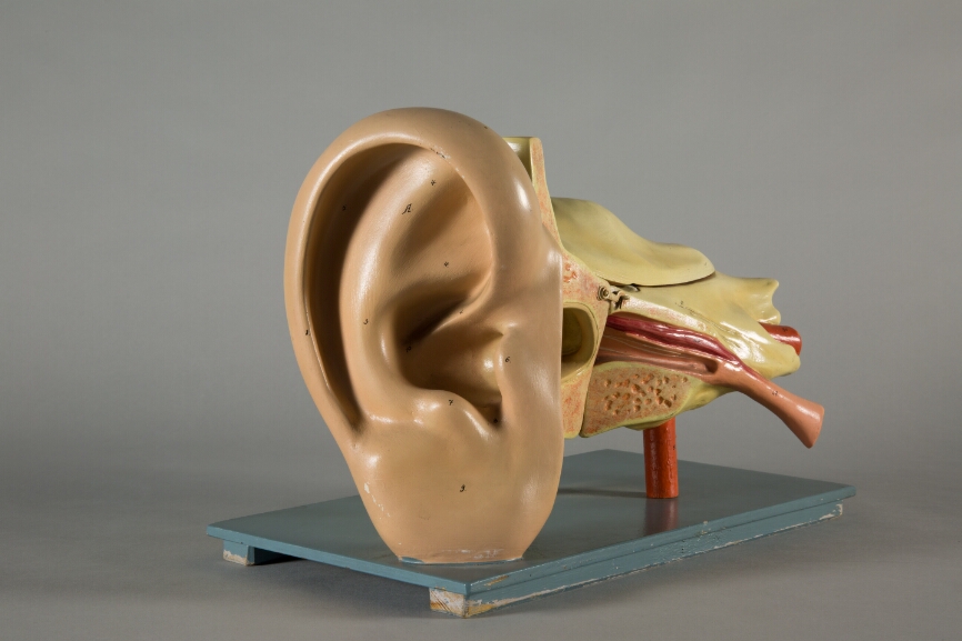
This is a model of a human ear. It can be taken apart completely. This way, you are able to carefully examine the internal structure of the ear. The model encompasses the outer ear, the middle ear as well as the inner ear.
How does the human ear function?
Together, the auricle and the external auditory canal make up the outer ear. It collects sound waves and directs them towards the eardrum, which causes it to vibrate. The vibrations are transferred onto the oval window via three small bones (hammer, anvil and stirrup) in the middle ear. The vibration of the oval window moves on through the fluid in the tubes of the cochlea. There, we encounter the auditory receptor cells. In turn, activation of these receptor cells triggers a biochemical reaction. This reaction stimulates the auditory nerve’s nerve cells, which finally activates the brain’s auditory cortex.

